

The rising prevalence of cardiovascular diseases, obesity, and a growing elderly population worldwide is driving the demand for minimally invasive diagnostic techniques that offer accurate diagnosis and faster recovery. According to Future Market Insights, the global angiography devices market is projected to grow from USD 14,343.8 million in 2025 to USD 26,176.4 million by 2035, registering a compound annual growth rate (CAGR) of 6.2%. CT and MRI angiography are among the most used non-invasive imaging techniques, selected based on the patient's clinical condition and the nature of the underlying pathology
CT-Angiography
CT angiography (CTA) involves the use of ionising radiation along with the injection of an iodinated contrast agent, typically administered through an intravenous line in the arm or hand. The CT scanner features a motorised X-ray source housed within a donut-shaped structure called the gantry.

During the procedure, the patient lies on a table that moves into the gantry, where the X-ray tube and detectors are positioned opposite to each other. As the gantry rotates around the body, X-rays pass through various tissues and are attenuated depending on their density. The detectors capture the exiting rays and transmit signals to a computer, which reconstructs high-resolution cross-sectional images using advanced algorithms.
While dense structures like bones are easily visualised, soft tissues such as blood vessels require contrast agents to enhance visibility. These agents improve X-ray attenuation, allowing for clearer imaging of vascular anatomy.
Clinically, CT angiography (CTA) is widely used to detect blood vessel abnormalities in the heart, brain, neck, chest, abdomen (such as kidneys and liver) and extremities, as well as in cases of trauma-related vascular injury. It plays a vital role in the early diagnosis of conditions such as stroke and myocardial infarction and is highly effective in identifying vascular abnormalities, including aneurysms, blockages, blood clots, malformations, trauma-related vessel injuries, and tumors, enabling timely and targeted clinical interventions.
Although there is a radiation risk in CTA, the amount of radiation exposure is low and controlled. As per risk analysis, the benefits of getting an accurate diagnosis via radiation outweigh the risks associated with radiation, which is completely acceptable. No radiation remains in your body after a CT scan.
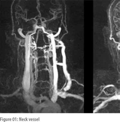
Proper precautions and preparation are necessary before taking a CTA scan since contrast agents may cause allergy to patients who have poor kidney function, and extra caution is required for pregnant women before scheduling the CTA scan.
MRI- Angiography
Magnetic Resonance Angiography (MRA) uses a powerful magnetic field and radiofrequency waves, along with advanced image reconstruction software, to evaluate blood vessels and detect vascular abnormalities. Unlike CT angiography, MRA does not involve ionising radiation. Gadolinium-based contrast agents are widely used in CE-MRA due to their positive paramagnetic substances, which increase the vascular signal by reducing the T1 relaxation time of blood.
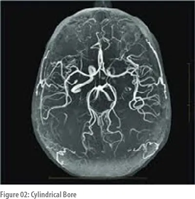
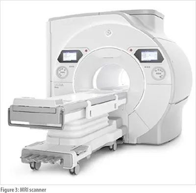
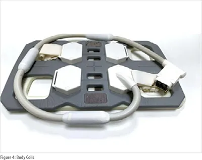
An MRI scanner consists of a large cylindrical bore surrounded by powerful magnets. The patient lies on a motorised table and is secured with straps and cushions to minimise movement and prevent blurry images. The scanner generates a strong magnetic field that causes hydrogen protons in the body to align with it. When a radiofrequency (RF) pulse is applied, these protons are temporarily displaced from their alignment. As they return to their original state, they release energy, which is detected by specialised MRI coils, such as head, body, or spine coils, depending on the area being scanned. This data is then processed by a computer to create detailed images of internal structures.
MRI is often able to tell the difference between diseased tissue and normal tissue better than X-ray, CT, and ultrasound. Contrast material used in MRI clearly define the blood vessels by making them appear bright white.
Individuals with certain metal implants, such as cochlear implants, pacemakers, or aneurysm clips, should not enter the MRI scanning area without prior safety assessments and precautions, as these devices may pose serious risks during the procedure. However, most orthopedic metal implants, such as screws or plates, are generally safe for MRI. That said, recently placed artificial joints may require alternative imaging methods depending on the material and timing of the surgery
Which is better?
Each imaging technique has its own advantages and limitations. Computed Tomography Angiography (CTA) offers excellent spatial resolution and a high signal-to-noise ratio, which helps differentiate small, closely packed structures, resulting in sharp and detailed images. However, due to the use of ionising radiation and iodinated contrast agents, CTA is typically reserved for patients with a higher likelihood of vascular pathology. In contrast, Magnetic Resonance Angiography (MRA) is a radiation-free imaging modality that employs various techniques to enhance image quality, making it a safer alternative for certain patient populations who need frequent scans for better treatment planning.
Recent advancements in CT and MRI Angiography
In the evolving landscape of angiographic imaging, technological advancements are focused on improving image quality, enhancing functional assessment, and integrating artificial intelligence (AI) for automated disease detection and better patient outcomes.
In CT angiography (CTA), innovations like photon-counting CT have significantly improved the ability to detect small structures in soft tissues, leading to higher diagnostic accuracy. Functional assessment tools such as CT-derived fractional flow reserve (CT-FFR) and intravascular imaging techniques like Optical Coherence Tomography (OCT) offer detailed insights into coronary physiology and plaque characteristics. These methods reduce the need for invasive catheter-based angiography and lower the risk of complications. Deep learning reconstruction (DLR) techniques further enhance vessel edge sharpness, reduce image noise, and improve pathology evaluation. Additionally, low-dose imaging protocols are increasingly adopted to minimise radiation exposure while maintaining excellent image quality, making CTA safer for a broader patient population.
MRI angiography has seen major improvements in both hardware and software. Modern 3T MRI scanners offer better signal-to-noise ratio and faster image acquisition, reducing motion artifacts and breath-hold requirements—enhancing patient comfort, especially in claustrophobic settings. High-V MRI systems improve imaging near metal implants and lungs. Due to concerns about gadolinium-related nephrogenic systemic fibrosis, there's growing interest in low-dose contrast-enhanced MRA (CE-MRA) and non-contrast MRA (NCE-MRA) techniques. Additionally, AI integration supports faster image reconstruction and workflow optimisation, improving diagnostic accuracy and reducing turnaround time.
Conclusion:
CT and MRI angiography continue to evolve with innovations that enhance diagnostic accuracy, safety, and patient comfort. CTA benefits from high-resolution imaging and functional tools, while MRA offers radiation-free alternatives with advanced contrast and AI integration. Together, these advancements are reshaping vascular imaging, making it more precise, accessible, and patient-friendly
References
1. SpringerLink Book - Sun Z, ed. Whole Body CT Angiography. Springer; 2024. doi:10.1007/978-981-97-1733-0
2. Medscape Article - Budoff MJ. Coronary CT Angiography. Medscape. https://emedicine.medscape.com/article/1603072-overview. Accessed September 13, 2025.
3. RSNA Journal Article - Flohr TG, et al. Dynamic CT angiography: principles and clinical applications. Radiographics. 2021;41(2):345-362. doi:10.1148/rg.210177
4. SpringerLink Book - Prince MR, ed. MR Angiography of the Body. Springer; 2023. doi:10.1007/978-3-540-79717-3
5. Radiology Key Chapter - Kinner S, et al. MR angiography techniques and clinical applications. In: RadiologyKey. https://radiologykey.com/mr-angiography-techniques-and-clinical-applications/. Accessed September 13, 2025.
6. International Journal Article - Lee JH, et al. Principles of non-contrast MR angiography techniques. Int J MRI. 2021;25(4):209-218. doi:10.13104/imri.2021.25.4.209
7. Source: https://www.ganeshdiagnostic.com/ct-angiography-neck-vessel