The Personalised Medicine from the Molecular Imaging Perspective
Personalising medical care to each patient's particular needs is referred to as customised medicine. Molecular imaging techniques are crucial in this regard. They play a big role in molecular features, long-term follow-up, disease heterogeneity assessment, detection, diagnosis, treatment, and planning for disease progression.
Introduction:
Cancer is one of the toughest issues in public health care due to its extensive effects and high mortality rates. This highlights the significance of advancing and supporting health care strategies and systems as well as the development of traditional medical approaches, such as chemotherapy, radiation therapy, and surgery, in order to eradicate cancer cells, improve patient survival rates, and ensure sustainability through the provision of adequate global healthcare based on prevention, precise diagnosis, and efficient therapy that has lower levels of multi-drug resistance, high effectiveness, and lower levels of cellular damage. The advent of customised medicine places a high priority on non-invasive imaging techniques for identifying diseases and treating patients, because they allow in vivo characterisation and measurement of important biomolecules and molecular events underlying malignant diseases, molecular imaging techniques have several benefits for enhancing clinical cancer care as well as drug discovery.
These imaging techniques can be classified into two categories: molecular imaging based on nanobodies and morphological/anatomical imaging. The development and application of molecular imaging has been facilitated by collaboration between specialists across a number of fields, including radiology, nuclear medicine, pharmacology, chemistry, molecular and cell biology, physics, mathematics, and engineering. Due to the widespread use of molecular imaging in treating numerous diseases, including cancer, in order to define the function of molecular imaging (ultrasound, MRI, PET, and SPECT) in personalised medicine, this text will analyse an extensive amount of published research on both topics. The article also explores the value of molecular imaging in the developing discipline of theranostics and how it might one day be used with other diagnostic methods to enhance the efficacy and efficiency of cancer treatment.
Ultrasound (US)
One of the most frequently utilised imaging modalities for diagnostic and clinical purposes is ultrasound, a high-resolution structural imaging tool. There is a lot of effort being made to increase the US's role in molecular imaging and personalised medicine. Some possible applications include adding sound pulses to MRI pulse sequences to increase the molecular field's use by US, improving drug delivery by speeding up lipid exchange and the propensity for fusion or improved contact between the nanoparticles and the targeted cell membrane while using the safe level of US waves, drug-loaded albumin-based carriers, and managing drug release.
Ultrasound Therapy
Additionally, researchers have strengthened contrast-enhanced ultrasounds to detect molecular markers and enhance ultrasound picture quality, making them suitable for ultrasound therapy. High-intensity frequency ultrasound treatment (HIFU), which induces hyperthermia or hypothermia, and thermal ablation are two highly effective approaches for thermal cancer therapy that use ultrasound as an imaging technology and biological system approach. To assess myocardial function, speckle tracking echocardiography (STE), a different ultrasound technique, is used. This method investigates the morphology of characteristic speckle patterns, natural myocardial acoustic indicators, during the cardiac cycle. Offline calculations of myocardial velocities and intrinsic heart deformation are possible (strain and strain rate). Even though it requires specialist software and requires greater image quality, this approach should be employed in patient follow-up.
Last but not least, a technique known as ultrasound elastography (USE) has shown the significance of tissue elasticity for the identification of malignancies. USE is a new instrument for evaluating tissue stiffness. During anti-stromal therapy and hyaluronic acid depletion, it tracks local changes in tissue pressure and assesses the spatial variance of the mechanical response. It is utilised as a non-invasive assessment of liver fibrosis in the clinical setting to characterise breast masses, assess thyroid nodules, target prostates that have undergone prostate biopsy, characterise focal renal lesions, and kidney and lymph node imaging are emerging.
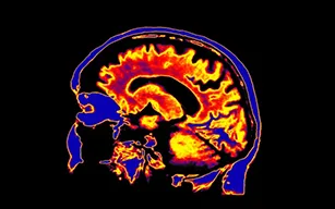
Magnetic Resonance Imaging (MRI)
One of the primary aspects of molecular imaging is the advancement of multimodality imaging, such as multiparametric magnetic resonance imaging (MRI) and PET/MRI, which are more routinely employed for detecting cancer, staging, and surveillance. This kind of imaging is becoming more vital because it helps doctors make diagnosis and plan treatments when inefficient surgeries and dangerous therapies could be avoided.
Based on the interaction of particular nuclei, typically protons, with molecules close to one another in intercellular tissue, magnetic resonance imaging (MRI) is a technique used to image the human body. A contrast is produced endogenously as a result of the different relaxation durations between different tissues. By selectively shortening the transverse T2 relaxation or the longitudinal T1 relaxation, external contrast agents might further enhance this.
Hybrid MRI
An important molecular marker employed in the in vivo imaging, characterisation, and quantification of biological activities in a tumour at both the cellular and molecular levels is fluorodeoxyglucose, the glucose used on a PET-MRI modality. When information on the morphology and function of a cancerous lesion are registered together, the innovation involved in combining these modalities improves the accuracy of the information. The ability to evaluate aberrant cellular signalling pathways in unprecedented depth is made possible when imaging and molecular diagnostics are combined. that is helped radiotracers and nuclear imaging tools are being created to track medicine effectiveness and advance the goal of customised treatment.
Single-Photon Emission Computerised Tomography (SPECT)
SPECT is using gamma rays with different energies to detect radiopharmaceuticals that offer sensitivity, the ability to quantify their uptake, and detection of these agents at any depth in the body, SPECT is one of the molecular imaging techniques that has really helped to visualise, characterise, and measure abnormal biologic processes of cancer at the macro and micro levels. This makes it appropriate for the monitoring of progress and outcome following surgery, radiotherapy, and other treatments. It might be feasible to select patients more carefully and advance the discovery, delivery, and development of new medications by developing personalised theragnostic compounds utilising the non-invasive imaging method known as SPECT.
Hybrid SPECT
Hybrid methods like SPECT/CT, PET/SPECT, and SPECT/MRI offer future development in molecular imaging by increasing sensitivity with molecular targeting, anatomic specificity, and resolution. The advantages of SPECT/CT include a short acquisition time of 1-2 min, minimal radiation exposure to the patient, and an excellent signal-to-noise ratio that permits the use of the scout picture for purposes other than attenuation correction. The personalised therapy and imaging of personal medicine are safe and suitable because these elements give the necessary pretherapy information on biodistribution, dosimetry, the limitation of vital organs or tissues, and the maximum tolerated dose. The features for separate modalities that improve the molecular and anatomical integration systems are SPECT/CT's main strength. Through better operational guidance, the avoidance of pointless procedures, and the provision of predictive data that helps guide change in both intra- and inter-modality therapy, SPECT/CT have a substantial impact on clinical management.
Positron Emission Tomography (PET)
PET is considered the gold standard in clinical molecular imaging. As a result, non-invasive detection down to the picomolar level is possible, because it has the great sensitivity needed for deep tissue penetration and the ability to see the majority of interactions between physiological targets and ligands, It has developed into the fastest-growing clinical imaging technology and is now used in cancer diagnosis and treatment planning due to its ability to quickly provide quantitative images and 3D morphological images, which enables dynamic imaging (time-resolved images to be generated).
Immuno-PET
Radiation therapy and immunotherapy together have the potential to revolutionise oncology, despite their promise not yet been completely realised. Immuno-PET imaging may be essential in supplying the essential data necessary to comprehend this complex link. Today, immuno-PET is a secure multimodality treatment approach that aids in the advancement of precision medicine. It does so by utilising radiolabelled antibodies and targets in combination with the high sensitivity and quantitative potential of PET non-invasively to provide quantitative, high quality, high spatial, and temporal resolution images that aid in estimating the antigenic expression level of immuno-PET, such as immune checkpoints and effector molecules, or the detection and tracking of cancer cells.
PET-CT
Due to its non-invasive nature and great accuracy in its use and management in oncology, PET-CT is the most extensively utilised and accessible molecular imaging technique. In addition to providing data on biodistribution, dosimetry, the limiting or crucial organ or tissue, and the maximum tolerated dose (MTD), PET-CT is a quantitative technology that offers information about morphologically relevant, physiological, and pathologic processes at the molecular level. It could measure abnormal molecular activity throughout the body and detect it, and it would be highly accurate at telling benign tumors from malignant ones. In order to facilitate simple management and early detection of tumor recurrences, it can also be utilised to assess the rates of chemotherapy response.
PET - US
PET-US uses radiolabelled microbubble shells with a short half-life, such as 18F-labeled, albumin-shelled, and VEGFR2-targeted, that are several micrometers in size. This technique provides improved quantification, which is especially true in biodistribution analysis, and can be utilised for targeted drug administration, such as administering VEGFR2 in breast cancer. It can be used to study the biodistribution of microbubbles following intravenous injection.
PET-MRI
PET-MR equipment is currently being developed and deployed in pre-clinical settings. High spatial resolution, temporal resolution, accuracy, enhanced soft tissue contrast, multi-planar capabilities, and reduced ionising radiation exposure are all made possible by this dual modality. These capabilities make it possible to conduct translational research from a cell culture setting to pre-clinical animal models to clinical applications, which is helpful for the drug discovery and evaluation process and could aid in optimising the non-invasive manufacturing of novel pharmaceuticals and the development of radiotracers.
PET OI
In recent years, PET and optical imaging have been combined and tested in vitro, ex vivo, or in vivo. The main advantages stem from the interaction of enhanced tissue penetration of radiation from positron emitter radionuclides, enabling non-invasive quantitative imaging and cancer detection and light produced by the fluorescent probe for optical imaging during surgery, especially robotic surgery. As a result, both the supplied dose and therapeutic efficacy can be properly tracked over time without producing systemic toxicity, enabling effective, tailored drug administration in vivo.
Conclusion
Personalised medicine attempts to improve diagnostic accuracy and lower therapy failure rates. Molecular imaging, a rapidly growing and fascinating field of study, is crucial to customised therapy. The long-term objective is for medical personnel to be able to use these approaches to improve diagnosis, improve treatment options, and predict patient outcomes, even if many parts of molecular imaging are still in their infancy. It is projected that in the coming decade, molecular imaging techniques will benefit from even more technological developments, which will ultimately have a substantial impact on customised medicine and lead to the emergence of a new eye for medicine.
References:
Salih, S.; Elliyanti, A.; Alkatheeri, A.; AlYafei, F.; Almarri, B.; Khan, H. The Role of Molecular Imaging in Personalized Medicine. J. Pers. Med. 2023, 13, 369. https://doi.org/10.3390/jpm13020369
Garra, B.S. Elastography: History, Principles, and Technique Comparison. Abdom. Imaging 2015, 40, 680–697.
Balber, T.; Tran, L.; Benˇcurová, K.; Raitanen, J.; Egger, G.; Mitterhauser, M. Experimental Nuclear Medicine Meets Tumor Biology.Pharmaceuticals 2022, 15, 227.
Cai, W.; Chen, X. Nanoplatforms for Targeted Molecular Imaging in Living Subjects. Small 2007, 3, 1840–1854.
Pysz, M.A.; Gambhir, S.S.; Willmann, J.K. Molecular Imaging: Current Status and Emerging Strategies. Clin. Radiol. 2010, 65, 500–516.
Belki´c, D.; Belki´c, K. Molecular Imaging in the Framework of Personalized Cancer Medicine. Isr. Med. Assoc. J. 2013, 15, 665–672.
Schillaci, O.; Urbano, N. Digital Pet/CT: A New Intriguing Chance for Clinical Nuclear Medicine and Personalized Molecular Imaging. Eur. J. Nucl. Med. Mol. Imaging 2019, 46, 1222–1225.


-(1).webp)